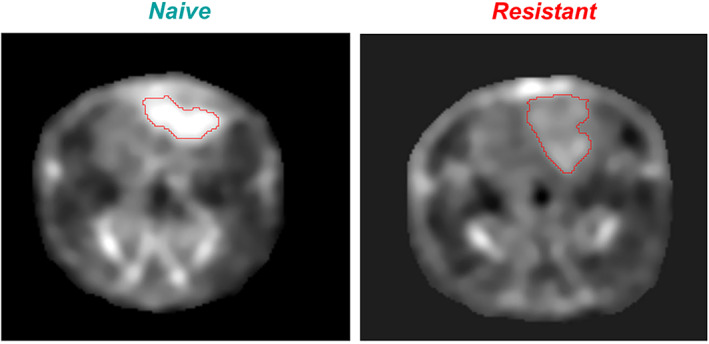
FIGURE 4.

Naive: sodium MRI of rat head with implanted naive glioma. The high concentration of sodium in glioma, visible as a bright spot on the image, indicates that tumor cells experience a large intracellular metabolic energy deficit and cannot maintain low intracellular sodium content, as seen in the normal brain around the glioma. These cancer cells are very sensitive to therapeutic interventions against tumors. The greater the sodium concentration in the tumor, the less resistant is the tumor. Resistant: sodium MRI of rat head with implanted resistant glioma (R1). The concentration of sodium inside the tumor is closer to the value in normal brain around the tumor. This small difference in sodium concentration indicates that the metabolic energy deficit is less dramatic in this tumor and they may have more cell density. Such tumor cells are more resistant to interventions, as is the case for normal cells around the tumor. Representative image reproduced from Ref. 55 with permission from John Wiley & Sons, NMR in Biomedicine.
