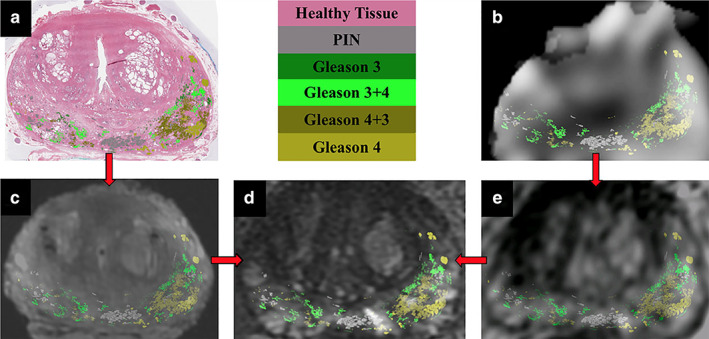
FIGURE 6.

Registration pipeline for all imaging data with Gleason contours overlaid. Whole‐mount histopathology (a) and the sodium‐MR image (b) are registered to the T2‐weighted ex vivo (c) and the lower‐resolution T2‐weighted in vivo images (e). The ex and in vivo images are individually registered to the high‐resolution T2‐weighted in vivo image (d). Gleason contour legends are shown in the upper middle panel. Figure reproduced from Ref. 80 with permission from John Wiley and Sons, Journal of Magnetic Resonance Imaging.
