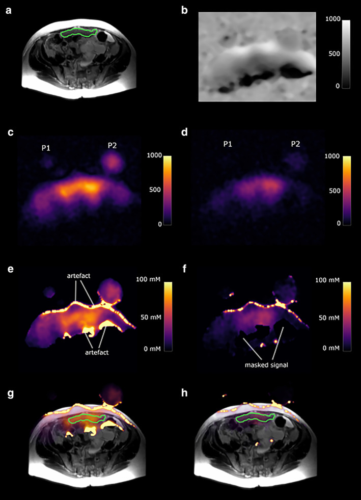
FIGURE 8.

A 73‐year‐old high‐grade serous ovarian cancer patient. P1 and P2 represent slices through the two sodium phantoms. The green outline shows a peritoneal cancer deposit. (a) T2‐weighted image. (b) Sodium B1 map; scale bar represents arbitrary units. (c) Total sodium image; scale bar represents image intensity. (d) Intracellular weighted sodium image; scale bar represents image intensity. (e) Masked total sodium concentration map; scale bar represents sodium concentration in mM. (f) Masked intracellular weighted sodium concentration map; scale bar represents sodium concentration in mM. (g) Fused T2W image and total sodium concentration map. (h) Fused T2W image and intracellular weighted sodium concentration map. Figure obtained from Ref. 31 with permission from Elsevier, European Journal of Radiology Open.
