
FIGURE 2.
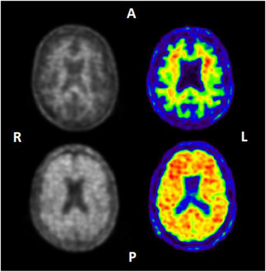
Example images of Neuraceq (left) and Vizamyl (right). Upper images: visually negative amyloid positron emission tomography (PET) scans showing only tracer retention in white matter. Lower images: visually positive amyloid PET scans showing tracer retention also in cortical areas. A, anterior; L, left hemisphere; P, posterior; R, right hemisphere