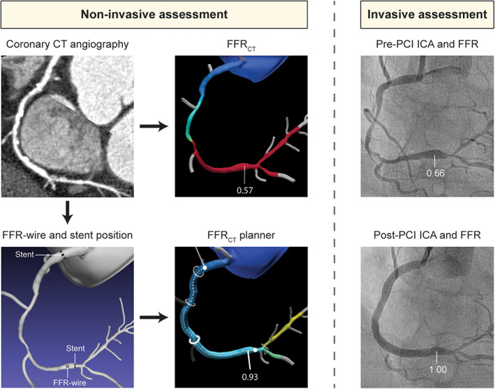
FIGURE 1.

Case example of non‐invasive assessment with FFRCT and FFRCT planner and invasive assessment with ICA and FFR in a patient undergoing revascularization. Non‐invasive coronary computed tomography (CT) angiography showed diffuse disease in the RCA, with multiple severe stenoses along the course of the vessel. FFRCT derived from standard coronary CT angiography images was calculated to be 0.57 in the distal RCA. Invasive assessment pre‐PCI confirmed diffuse disease in the mid and distal RCA with a corresponding FFR in the distal RCA of 0.66. Subsequently, PCI was performed with implantation of three stents with a total stent length of 81 mm, resulting in a post‐PCI FFR of 1.00. For computation of FFRCT planner, the location of invasive FFR measurement and actual stent location were annotated in a computational model by a researcher blinded to invasive data. After simulation of stenosis removal, FFRCT planner value was shown to 0.93. FFR, fractional flow reserve; FFRCT, computed tomography derived FFR; ICA, invasive coronary angiography; PCI, percutaneous coronary intervention [Color figure can be viewed at wileyonlinelibrary.com]
