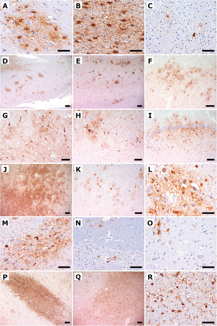
Fig. 2.

Immunohistochemical findings of hyperphosphorylated tau using the antibody AT8. (A) The cingulate gyrus shows a cluster of GFA tau deposits. (B) The temporal ccortex shows severe white matter ARTAG. (C) The temporal white matter shows focal granular astroglial tau deposits. (D) The cingulate gyrus shows patchy clustering of GFAs in the grey matter and additionally white matter ARTAG. (E) The temporal cortex shows grey matter ARTAG. (F) The ambient gyrus shows grey matter ARTAG. (G) CA1 shows neuronal tau pathology, including NFTs, pretangles, neuropil threads, and additional of grey matter ARTAG. (H) The CA2/3 sectors show GFAs and neuronal tau pathology. (I) The dentate gyrus shows prominent grey matter ARTAG. (J) The amygdala shows extensive grey matter ARTAG. (K) The accumbens nucleus shows patchy clusters of GFA. (L) The substantia nigra shows pronounced astrocytic tau pathology. (M) The locus ceruleus shows neuronal and more astroglial tau pathology. (N) Pontine base shows a single NFT and astroglial tau immunoreactivity. (O) The dentate nucleus shows astroglial tau immunoreactivity. (P) The temporal white matter shows extensive white matter ARTAG as well as perivascular ARTAG. (Q) The amygdala shows severe white matter ARTAG including perivascular ARTAG. (R) The pyramidal tract shows white matter ARTAG. Scale bars: 200 μm (D, E, J, P, Q), 100 μm (F‐I, K, M, N), 50 μm (A‐C, L, O, R).
