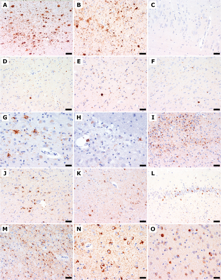
Fig. 3.

Immunohistochemical findings of α‐synuclein (A–F), phosphorylated TDP‐43 (G–N), and TDP‐43 (O). (A) The locus coeruleus shows Lewy bodies, Lewy neurites, and diffuse granular cytoplasmic immunoreactivity. (B) The amygdala shows Lewy bodies, Lewy neurites, and astroglial α‐synuclein immunoreactivity. (C) The CA2/3 sectors show single delicate dot‐like α‐synuclein‐immunoreactive neurites. (D‐F) The entorhinal cortex (D), putamen (E), and gyrus cinguli (F) show Lewy bodies and Lewy neurites. (G) The inferior olive shows phosphorylated TDP‐43 immunoreactivity with astroglial cytoplasmic deposits and threads. (H) The pontine nucleus shows astroglial cytoplasmic phosphorylated TDP‐43 immunoreactivity and fine threads. (I) The septum nucleus shows marked diffuse neuronal pathology and fine threads and granular deposits in the neuropil. (J) The cingulate gyrus shows thread pathology, neuronal and astroglial phosphorylated TDP‐43 immunoreactivity. (K) The eEntorhinal cortex shows thread pathology and neuronal and astrocytic phosphorylated TDP‐43 immunoreactivity. (L) The dentate gyrus shows threads, and compact, neuronal, and phosphorylated TDP‐43 immunoreactivity. (M) The accumbens nucleus shows marked diffuse cytoplasmic neuronal pathology and fine threads and granular neuropil deposits. (N) The amygdala shows severe neuronal and here only few astrocytic cytoplasmic immunoreactivity as well as abundant fine threads. (O) The amygdala shows loss of neuronal nuclear physiological TDP‐43 immunoreactivity and compact cytoplasmic inclusions. Scale bars: 100 μm (A); 50 μm (B‐F, I‐M); 25 μm (G, H, N, O).
