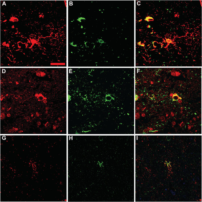
Fig. 4.

Findings of doube immunofluorescence staining in the brain. (A–C) Double immunofluorescence of the amygdala using antibodies against GFAP (red, A) and hyperphosphorylated tau (green, B) reveals accumulation of hyperphosphorylated tau in astrocytes (yellow, C). (D–F) Double immunofluorescence staining of the amygdala using antibodies against TDP‐43 (red, D) and hyperphosphorylated tau (green, E) shows codistribution of both proteins within the cytoplasm of some cells with neuronal morphology (yellow, F). (G–I) Double immunofluorescence staining of the entorhinal cortex using antibodies against phosphorylated TDP‐43 (green, H) and hyperphosphorylated tau (red, G) shows codistribution of both proteins within the cytoplasm of some cells with astroglial morphology (yellow, I). Scale bar: 10 μm (A‐I).
