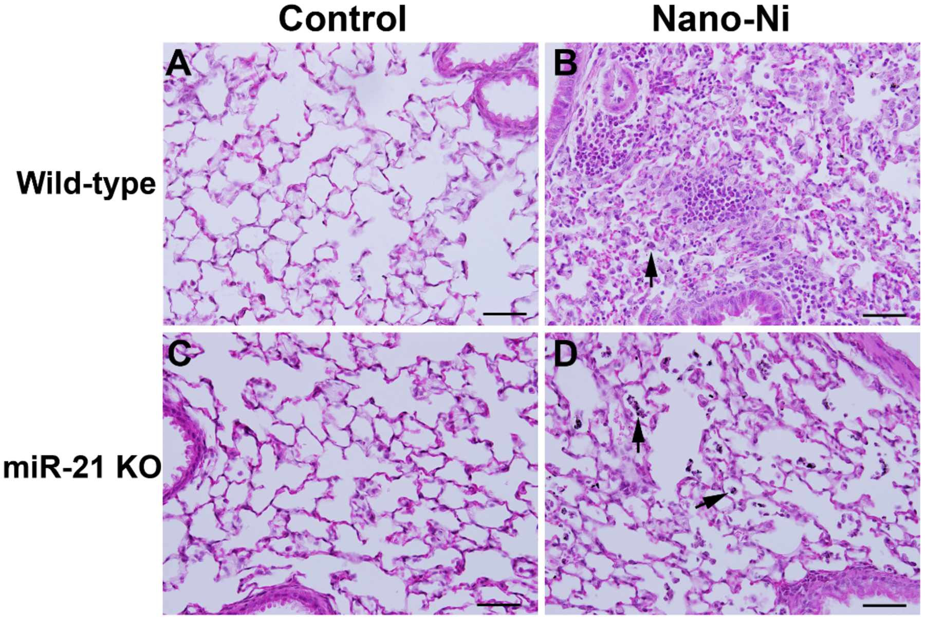
Figure 4.

Acute pulmonary inflammation in mouse lungs at day 3 after Nano-Ni exposure. Wild-type and miR-21 KO mice were instilled intratracheally with 50 μg per mouse of Nano-Ni. Control mice were instilled with physiological saline. Lung tissues collected from mice at day 3 after exposure were analyzed by H&E staining. A and C show the normal structure of alveoli and peribronchiolar areas in the control mice. B shows acute inflammation in the lungs of a wild-type mouse with Nano-Ni exposure, evidenced by large numbers of polymorphonuclear (PMN) cells and macrophages infiltrating into the lung parenchyma. Acute pulmonary inflammation was also observed in the lungs of Nano-Ni-exposed miR-21 KO mice, but to a much lesser extent (D). Arrows: Nano-Ni-phagocytized macrophages. Scale bar represents 50 μm in all panels.
