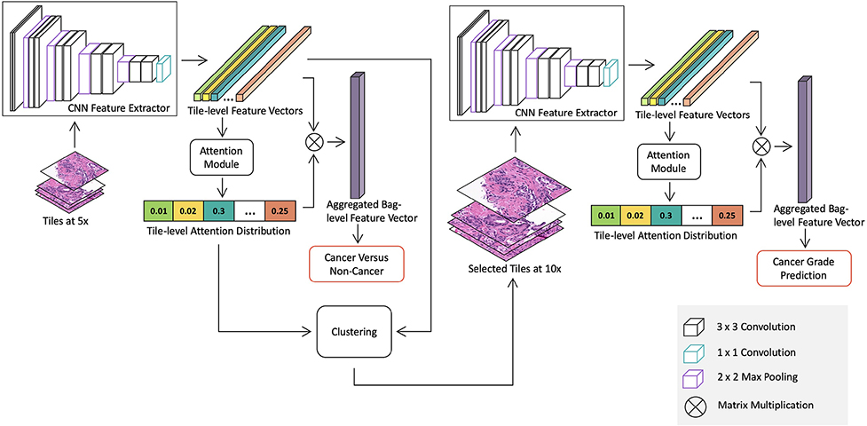
Figure 1:
Overview of the proposed whole slide image detection and classification model. The model consists of two stages: a cancer detection stage at a low magnification and a cancer classification stage at a higher magnification for suspicious regions. Both stages contains a CNN feature extractor, which is trained in the MIL framework with slide-level labels. Specifically, the detection stage model is trained with all tiles extracted from slides at 5x to differentiate between benign and malignant slides. The attention module in the detection stage model produces a saliency map, which represents relative importance of each tile for predicting slide-level labels. Then we use the K-means clustering method to group tiles into clusters based on tile-level features. The number of tiles selected from each cluster is determined by the mean of cluster attention values. Discriminative tiles identified by the detection stage model are then extracted at 10x and fed into the classification stage model for cancer grade classification.

