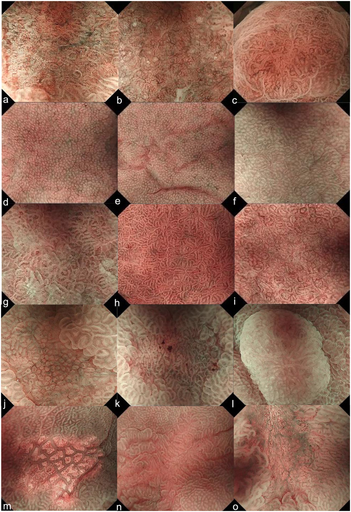
Figure 1.

Educational endoscopic images used for the CNN. (a) Differentiated‐type cancer (0‐IIc, tub1), (b) differentiated‐type cancer (0‐IIc, tub2), (c) differentiated‐type cancer (0‐IIa, tub1), (d–f) fundic gland mucosa, (g–i) pyloric gland mucosa, (j, k) patchy redness, (l) adenoma, (m) xanthoma, (n) focal atrophy, and (o) ulcer scar. 0‐IIa, flatly elevated; 0‐IIc, flatly depressed; CNN, convolution neural network; tub1, well differentiated adenocarcinoma; tub2, moderately differentiated adenocarcinoma.
