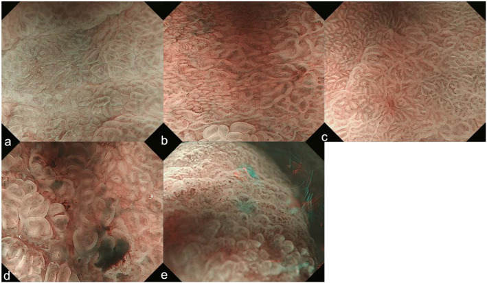
Figure 2.

Misdiagnosed endoscopic images shown by the CNN. (a–c) False negatives: lesions appear as intestinal metaplasia or gastritis. (a) Differentiated‐type cancer (0‐IIc, tub1, after Hp eradication): regular MVP + regular MSP with a DL. (b) Differentiated‐type cancer (0‐IIc, tub2, after Hp eradication): regular MVP + regular MSP with a DL. (c) Differentiated‐type cancer (0‐IIa, tub1, after Hp eradication): regular MVP + regular MSP with a DL. (d) False negative: bleeding. (e) False negative: low‐power view and out of focus. 0‐IIc, flatly depressed; CNN, convolution neural network; DL, demarcation line; Hp, Helicobacter pylori; MSP, microsurface pattern; MVP, microvascular pattern; tub1, well differentiated adenocarcinoma; tub2, moderately differentiated adenocarcinoma.
