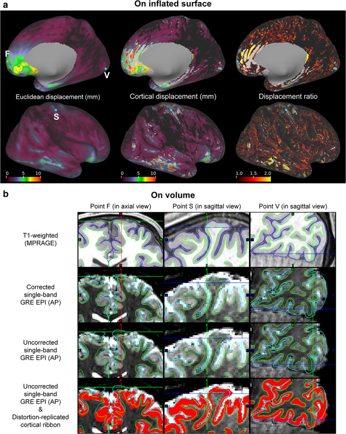
FIGURE 8.

Individual effects of distortion corrections on cortical surface‐based analysis. (a) Maps of Euclidean (left column) and cortical displacements (middle column), and displacement ratio (right column) are shown on the right inflated surface of a typical subject. These maps from medial (top row) and lateral views (bottom row) are aligned. (b) Cross‐sectional views of the points F, S, and V in (a) are shown in the left, center, and right columns, respectively. Crosshairs correspond to these points. In each cross‐sectional view, T1‐weighted and corrected single‐band (SB) gradient‐echo (GRE) echo‐planar images are shown in the first and second rows, respectively. The uncorrected versions of the echo‐planar images are also shown in the third row. The distortion‐replicated cortical ribbon denoted in red is overlaid on the uncorrected images in the fourth row. Green and blue or aqua lines indicate pial and white surfaces created with FreeSurfer, respectively.
