
Figure 2.
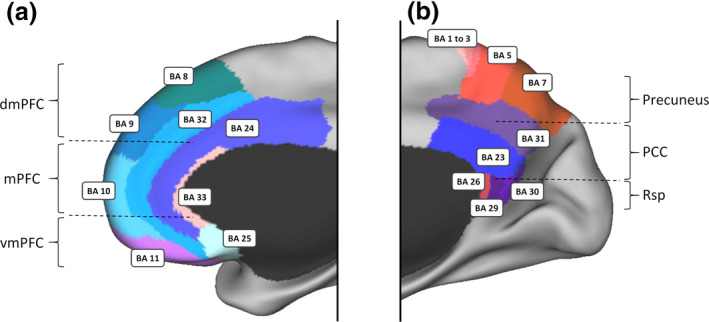
Anatomy of the midline DN core based on Brodmann cytoarchitecture. Panel (a) illustrates the approximate location of the mPFC, which is often described as primarily comprising areas 32, 24, and 25 (Córcoles‐Parada et al., 2017). However, recent parcellations of the DN consider that the mPFC can be subdivided into three parts (dmPFC, mPFC, and vmPFC) with only the mPFC being part of the DN midline core. The dmPFC is more strongly associated with regions involved in mentalizing and social cognition, while the vmPFC is more closely related to limbic and subcortical areas supporting motivational and valuation processes (Andrews‐Hanna et al., 2010; D’Argembeau, 2013; Yeo et al., 2011). Areas 12 and 14 are not represented as they were not explicitly defined in humans by Brodmann (1909). Panel (b) illustrates the approximate location of the PCC, which was initially considered as consisting of areas 23, 29, 30, and 31 (Brodmann, 1909). However, more recent views consider that the PCC only includes the posterior parts of areas 23 and 31 (Leech & Sharp, 2014; Vogt, 2009) with areas 29 and 30 being parts of the Rsp. Area 7 is usually referred to as the Precuneus, while areas 1, 2, 3, and 5 are parts of the sensory‐motor system. dmPFC, dorsal medial prefrontal cortex; mPFC, medial prefrontal cortex; vmPFC, ventral medial prefrontal cortex; PCC, posterior cingulate cortex; Rsp, retrosplenial cortex. The Brodmann areas are displayed on the PALS‐B21 inflated surface map using CARET software (Van Essen, 2005).