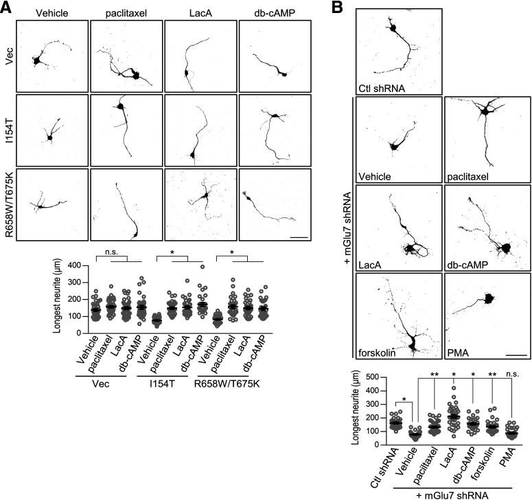
Figure 7.
The impaired axon outgrowth by mGlu7 variants or knock-down is restored by adjusting dysregulated cytoskeletal dynamics or cAMP signaling. A, Cortical neurons (DIV1) were cotransfected with EGFP and control vector (Vec), mGlu7 I154T, or R658W/T675K rescue construct. Twenty-four hours after transfection, the neurons were treated with the indicated reagents for 24 h. At DIV3, axonal outgrowth was visualized by confocal microscopy and measured by NeuronJ software. Scale bar: 50 μm. Scatter plots represent the mean ± SEM, n.s., not significant, *p < 0.0001. B, Hippocampal neurons (DIV1) were transfected with lentiviral vector harboring control (Ctl) shRNA or mGlu7 shRNA. Twenty-four hours after transfection, the neurons were treated with the indicated reagents for 24 h. Scale bar: 50 μm. Scatter plots represent the mean ± SEM, n.s., not significant, *p < 0.0001, **p = 0.0002.

