Figure 8.
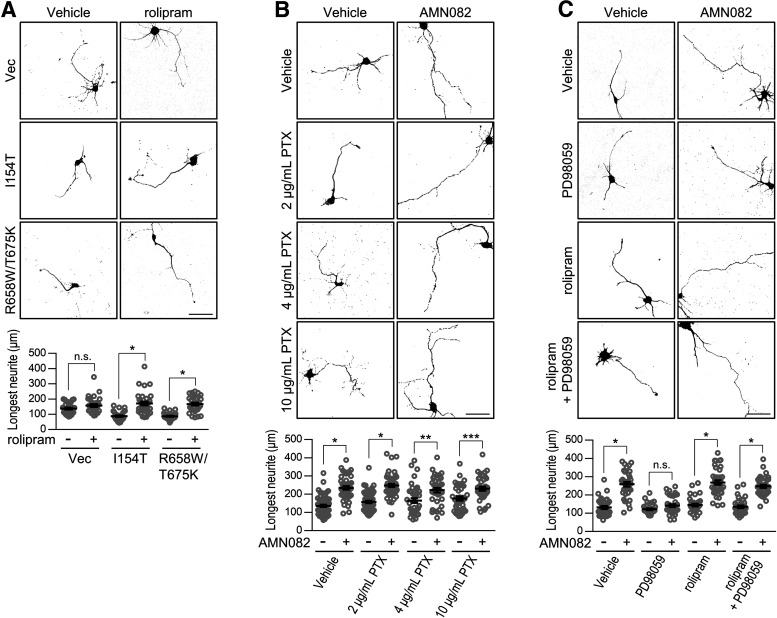
mGlu7-mediated axon outgrowth is regulated by the MAPK-cAMP pathway, but is independent of the Gαi pathway. A, Cortical neurons (DIV1) were cotransfected with EGFP and the control vector (Vec), mGlu7 I154T or R658W/T675K rescue constructs in the absence or presence of rolipram (500 nm). At DIV3, axonal outgrowth was visualized by confocal microscopy and measured by NeuronJ software. Scale bar: 50 μm. Scatter plots represent the mean ± SEM, n.s., not significant, *p < 0.0001. B, mGlu7-mediated axon outgrowth is not related to the Gαi signaling pathway. After cortical neurons (DIV1) were transfected with EGFP, the neurons were treated with AMN082 in the presence or absence of 2, 4, or 10 μg/ml PTX, an inhibitor of Gα subunits of the heterotrimeric G-protein. At DIV3, axonal outgrowth was visualized by confocal microscopy. Scale bar: 50 μm. Scatter plots represent the mean ± SEM. *p < 0.0001, **p = 0.0041, ***p = 0.0085. C, MAPK is an upstream mediator of cAMP in mGlu7-mediated axon outgrowth. After transfection with EGFP, the cortical neurons (DIV1) were treated with MEK inhibitor (PD98059) and/or 500 nm rolipram, a PDE inhibitor that suppresses cAMP degradation, in the absence or presence of the mGlu7 agonist AMN082. At DIV3, axonal outgrowth was visualized by confocal microscopy. Scale bar: 50 μm. Scatter plots represent the mean ± SEM, n.s., not significant, *p < 0.0001.

