Figure 3.
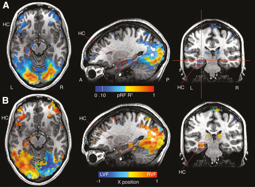
Retinotopic sensitivity in the hippocampus is spatially separate from PHG. A, pRF R2 is overlaid onto axial, sagittal, and coronal slices of a representative participant. Strong responses are evident throughout visual cortex and extend anteriorly in ventral temporal cortex. Two clusters within the hippocampus (red boxes) appear spatially distinct from more posterior responses in PHG. B, The x-position of pRF centers are overlaid onto the same slices. The two hippocampal clusters show pRF positions firmly in the contralateral (right) visual field.

