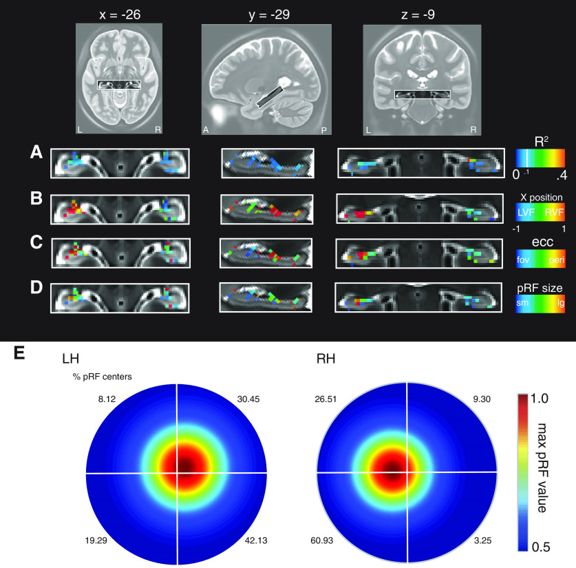
Figure 5.
pRF parameters in the hippocampus from the HCP data. A, Enlarged axial, sagittal, and coronal views of the hippocampus shown with the pRF R2 overlaid. Voxels in the hippocampus are well fitted by the pRF model, with clusters in anterior medial portions. B, The x-position of pRFs is shown. In general, pRFs show largely contralateral visual field positions. C, pRF eccentricity suggests peripheral pRFs in the hippocampus. D, Hippocampal pRFs appear also to be large. E, Visual field coverage from all suprathreshold pRFs (R2 > 0.1; left = 199, right = 115). A contralateral bias is present in bilateral hippocampus. The percentage of pRF centers in each quadrant are inset.

