
Figure 1. Deficiency of Runx1 in chondrocytes and osteoblasts caused the dwarfism, decreased body weight, and decreased bone density in mutant mice.
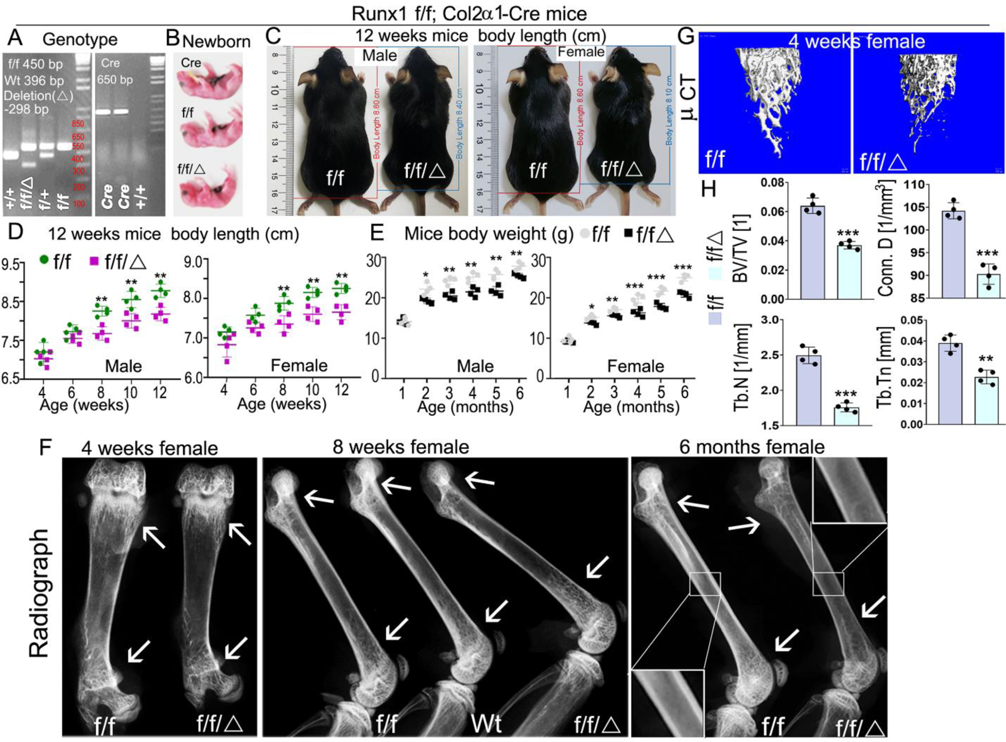
(A) PCR genotyping images of Runx1 f/f and Runx1f/fCol2α1-cre mice. f, Runx1 allele carrying loxP sites; +, WT Runx1 allele; Δ, Runx1 allele with loxP floxed sequence deleted. (B) Gross appearances of newborn Runx1f/fCol2α1-cre mice and littermates. (C) Measurement of mice body lengths from 12-week-old Runx1f/fCol2α1-cre and Runx1f/f mice. (D) Quantification of mouse body lengths from 4-weeks-old to 12-weeks-old. (E) Quantification of mouse body weights from 1-month-old to 6-months-old. (F) X-ray analysis long bones from 4-week, 8-week, and 6-month-old Runx1f/fCol2α1-cre mice and littermate controls (f/f), white arrows referred to cortical bone and trabecular bone density differences between wt and mutant mice. (G) Micro computed tomography (μCT) examination was performed to determine the bone formation in 4-week murine femurs. (H) Quantification data of bone volume/tissue volume (BV/TV), the trabecular bone numbers (Tb. N), trabecular bone thickness (Tb. Tn), and connectivity density (Conn. D) in G. All data are presented as mean ± SD, n=4, NS denotes not significant, *p<0.05, **p<0.01, ***p<0.001. f/f denotes Runx1f/f; f/f/Δ denotes Runx1f/fCol2α1-cre.