
Figure 2. Postnatal Runx1f/fCol2α1-cre mice display impaired trabecular bone formation and deformed growth plates.
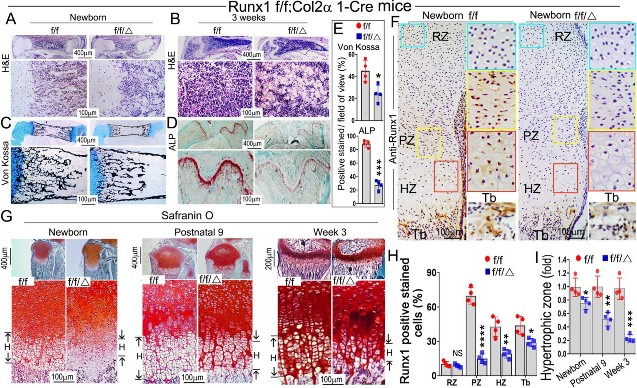
(A, B) Hematoxylin and eosin (H&E) staining of (A) newborn and (B) 3-week-old murine Runx1f/fCol2α1-cre and Runx1f/f femurs. (C) Von Kossa staining of the newborn murine femurs. (D) ALP staining of 3-week-old murine femurs and tibias. (E) Quantification of (C) Von Kossa staining and (D) ALP staining. (F) Immunohistochemistry (IHC) staining of newborn wild type and Runx1f/fCol2α1-cre mice femur sections by using anti-Runx1 antibodies to detect the expression of Runx1 in the growth plates. RZ, resting zone; PZ, proliferation zone; HZ, hypertrophic zone; Tb, trabecular bone. (G) Safranin O staining of the tibias from newborn, postnatal 9, and 3-week old mice. H denotes hypertrophic zone. (H) Quantification of Runx1 positive staining cells in (F). (I) Measurement the length of the hypertrophic zone in (G). All data are presented as mean ± SD, n=6, NS denotes not significant, *p<0.05, **p<0.01, ***p<0.001.