
Figure 6. Runx1 may interfere with Ihh signaling to regulate chondrocyte proliferation and differentiation in vitro.
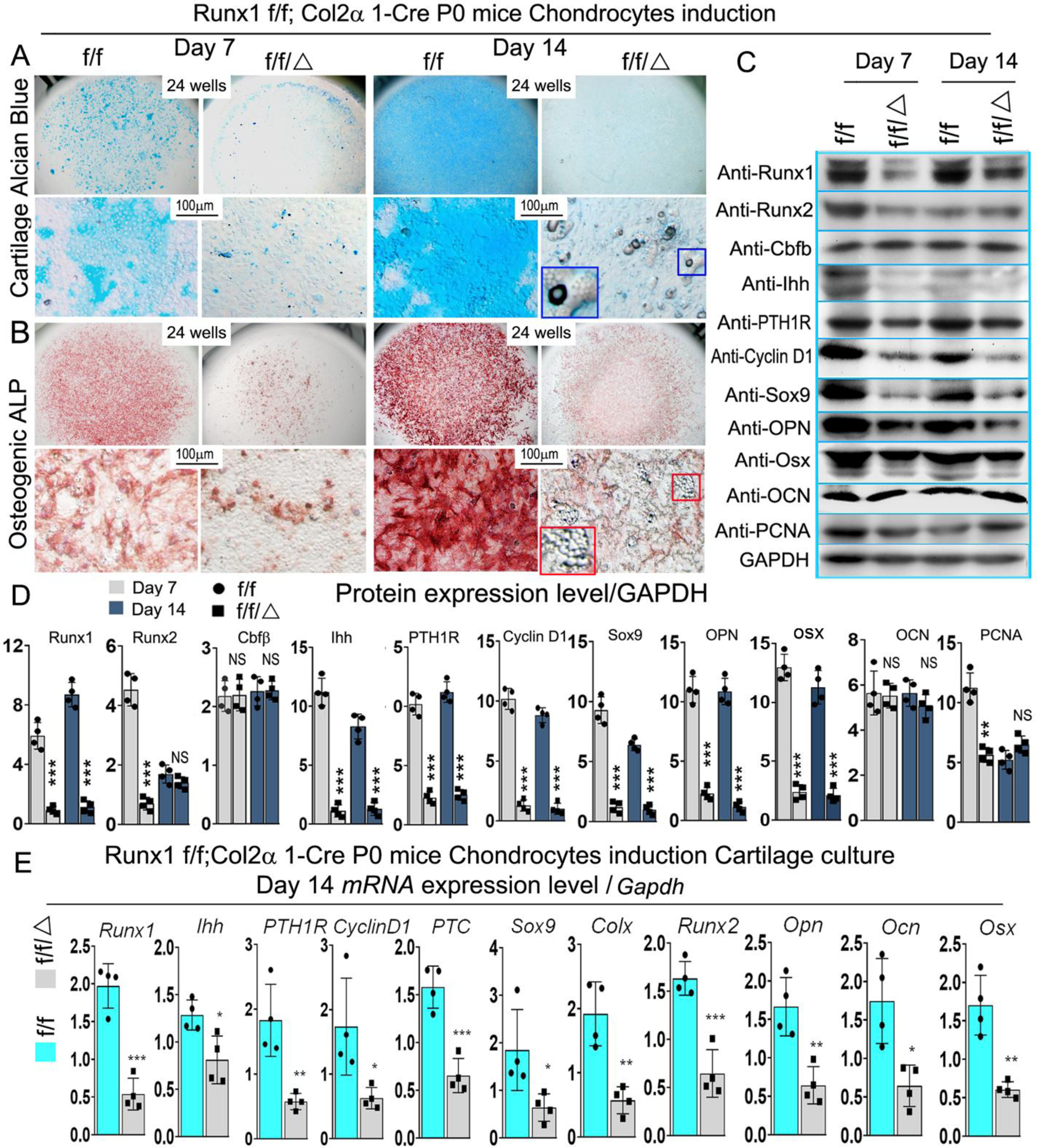
(A, B) Micromass culture of chondrocytes derived from Runx1f/fCol2α1-cre and Runx1f/f newborn mice growth plates. The blue and red boxed and zoomed area reffered to adipocytes. (A) Alcian blue and (B) ALP staining was performed at day 7 and day 14. (C) Western blot was performed on cultured chondrocytes of day 7 and day 14 from A to evaluate the protein levels of Runx1, Runx2, Cbfβ, Ihh, PTH1R, Cyclin D1, Sox9, Opn, Osx, Ocn, and PCNA. (D) Quantification data of (C). (E) qPCR was used to analysis the expression level of Ihh signaling related genes in day 14 chondrocytes. All data are presented as mean ± SD, n=4, NS denotes not significant, *p<0.05, **p<0.01, ***p<0.001.