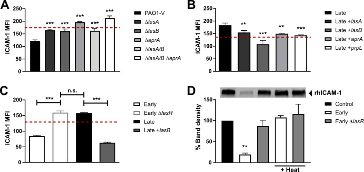
Fig 3. LasR-regulated proteases degrade ICAM-1.
BEAS-2B cells were stimulated for 24h with 30 μL filtrates of (A) PAO1-V and its isogenic protease mutants of lasA, lasB and aprA, (B) the Late isolate and complemented strains Late +lasA, Late +lasB, Late +aprA or Late +prpL (T4P) or (C) Early, Early ΔlasR, Late, Late +lasB. mICAM-1 levels were measured by flow cytometry, with SCFM (media) serving as negative control (—dashed line). (D) in vitro degradation of recombinant human ICAM-1 (rhICAM-1) by P. aeruginosa filtrate. In each sample, 250 ng rhICAM-1 was incubated with 5 μL filtrates of the Early and Early ΔlasR strains (+/- heat inactivation) or PBS control for 24h, and the remaining intact rhICAM-1 following degradation was quantified by Western Blotting. Results in (A), (B) and (C) are shown as mean ± SD from one representative experiment (from ≥ 2 independent experiments, each with biological triplicates). Results in (D) are displayed as a representative Western Blot and quantification of the % band density compared to the PBS control condition (n ≥ 3 biological replicates from two independent experiments). Different lanes were cropped from the same blot and imaged at the same exposure. Full blots can be found in the S1 Spreadsheet. *P < 0.05; **P < 0.01; ***P < 0.001.

