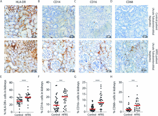
Fig 3. Increased numbers of CD14+ and CD16+ cells are detected in kidneys during HFRS.
(A-C) Representative images of HRP-based kidney immunohistochemistry for (A) HLA-DR, (B) CD14 and (C) CD16 and (D) CD68 in control (top panels) and acute HFRS patients (lower panels), both patients diagnosed with acute tubulointerstitial nephritis. Hematoxylin was used as the counterstain. (E-H) Graphs show percentages of (E) HLA-DR (F) CD14 (G) CD16 and (H) CD68 positive cells out of all cells in 43 non-HFRS controls and 37 acute HFRS kidney tissues as assessed by automated computing methods. Statistical differences between groups were assessed by Mann-Whitney test and considered significant at p<0.05. (***p <0.001 and ****p< 0.0001).

