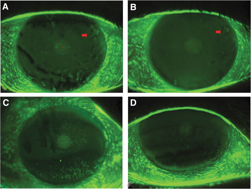
FIG. 1.
Fluorescein staining of the ocular surface. Please note the evident reduction of corneal staining after 28 days of treatment (B, D) versus baseline (A, C). The arrow indicates the presence of an alteration of the corneal epithelial basement membrane at baseline (A) and after 28 days of treatment (B).

