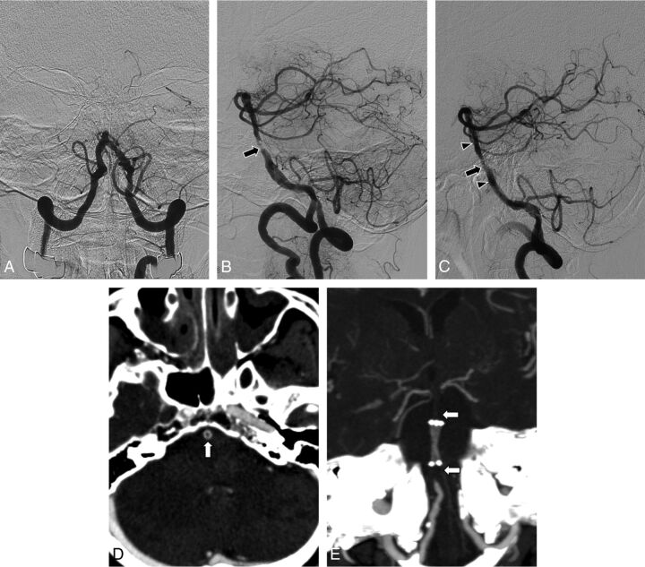
Fig 1.
Brain images obtained in a patient with acute stroke. A, Anteroposterior view of a vertebral angiogram shows arterial occlusion at the proximal portion of the basilar artery. B, Lateral projection of the vertebral angiogram obtained after stent-retriever embolectomy reveals severe underlying stenosis (arrow) at the site of arterial occlusion. There were no retrieved thrombi with the Solitaire stent. C, Lateral projection of vertebral angiography performed after intracranial stent placement shows suboptimal angioplasty, with residual stenosis of 60% (arrow). Arrowheads indicate the proximal and distal ends of the stent. D, Axial source image of CTA shows hypoattenuation (arrow) within the stented segment of the basilar artery. E, Maximum-intensity-projection image of the follow-up CTA 2 days after the procedure shows a discontinuation of the contrast column both proximal and distal to the stent (arrows).

