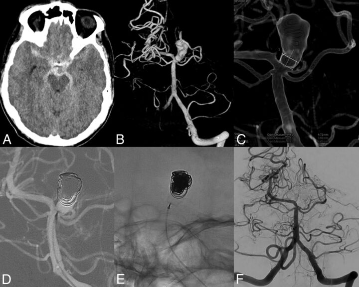
Fig 2.
A 61-year-old man with a ruptured basilar tip aneurysm treated with the WEB and coils. A, CT scan with subarachnoid blood. B, 3D angiogram reveals a 13-mm basilar tip aneurysm. C, Neck measurement on a 3D angiogram. D and E, After placement of coils in the dome and a WEB-SL, 6 × 3 mm, in the neck. F, Three-month follow-up angiogram with complete occlusion.

