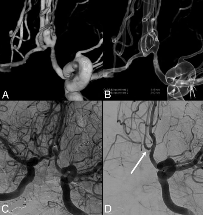
Fig 3.
A 57-year-old woman with a ruptured anterior communicating artery aneurysm. A, 3D angiogram shows a small anterior communicating artery aneurysm. Note the spasm in the left A1. B, Measurement of the height (3.9 mm) and neck width (2.3 mm). C, Angiogram directly after placement of a 4-mm WEB-SLS. Note some opacification inside the WEB. D, Angiogram at 3 months demonstrates complete occlusion of the aneurysm.

