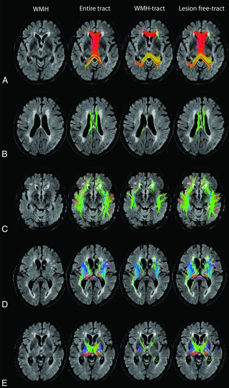
Fig 1.

Tract segmentation, WMH tracts, and lesion-free tracts. Axial T2 FLAIR demonstrates white matter hyperintensities, tractography representation of the entire tracts, WMH tracts, and lesion-free tracts in the corpus callosum (A), cingulum (B), association fibers (C), corticospinal/bulbar tract (D), and thalamic tracts (E).
