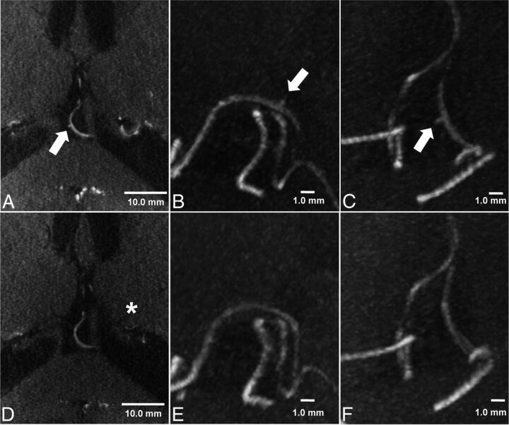
Fig 3.
TOF-MRA and MIP of TOF-MRA at 7T before (A–C) and 6 months after (D–F) the bypass operation in case 5. A microaneurysm (560-μm maximum diameter) is delineated arising from a collateral vessel of the posterior choroidal artery in the third ventricle (arrow). Six months after the bypass operation, MIP of TOF-MRA shows disappearance of the microaneurysm. The signal intensity of collateral vessels has decreased in the matched axial planes (asterisk). The scale bar indicates 10.0 mm (TOF-MRA, A and D) and 1.0 mm (MIP image, B and C; E and F), respectively.

