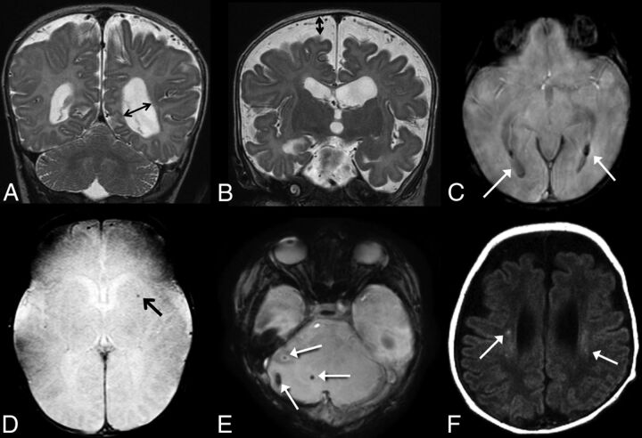
Fig 2.
A, Coronal T2-weighted image demonstrates ventriculomegaly, scored as grade 2 on our scale; B, Coronal T2-weighted image demonstrates enlarged extra-axial spaces, scored as grade 2 on our scale. C, SWI shows evidence of grade 2 intraventricluar hemorrhage (arrows). D, SWI shows evidence of parenchymal hemorrhage, grade 1 on our scale (arrow). E, SWI shows foci of cerebellar hemorrhage, grade 2 on our scale (arrows). F, Axial T1 weighted images demonstrate foci of bilateral white matter injury, grade 2 on our scale (arrows).

