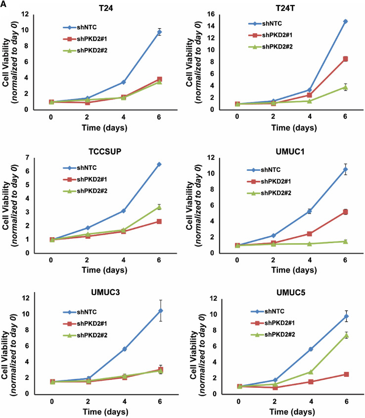
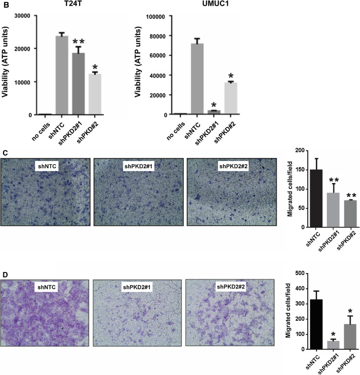
Fig. 8.
Knockdown of PKD2 expression decreases proliferation, anchorage-independent growth, and invasion of bladder carcinoma cells in vitro. a Six cell lines (T24, T24T, TCCSUP, UMUC1, UMUC3, and UMUC5) were transduced with lentivirus coding for human shRNA sequences for PKD2 (shPKD2#1 and shPKD2#2) or for the control (shNTC). Cell growth and proliferation were assessed by MTT assay at days 0, 2, 4, and 6 after cell plating. P < 0.01 for the shPKD2 groups vs. the control shNTC group at day 6 in all the six cell lines tested. b Depletion of PKD2 abrogates bladder cancer cell growth in low attachment. T24T and UMUC1 cell lines were transduced with lentivirus coding for human shRNA sequences for PKD2 (shPKD2#1 and shPKD2#2) or for the control (shNTC). Cell growth in low attachment was assessed by GILA assay at day 5 after cell plating, as described in “Materials and methods”. *P < 0.01 and **P < 0.05 for the shPKD2 groups vs. the control shNTC group. c, d Knockdown of PKD2 expression compromises the invasive ability of bladder carcinoma cells. TCCSUP cells (c) and UMUC1 cells (d) were transduced with lentivirus coding for human shRNA sequences for PKD2 (shPKD2#1 and shPKD2#2) or for the control (shNTC). A two-chamber cell invasion system was set up, as described in “Materials and methods”, and the number of invading cells was counted in ten random microscopic fields (×40). The numeric data for the two cell lines are shown in the two column graphs, respectively. Values are expressed as mean ± SD. **P < 0.05 and *P < 0.01 for the shPKD2 groups vs. the shNTC group in TCCSUP and UMUC1 cells, respectively


