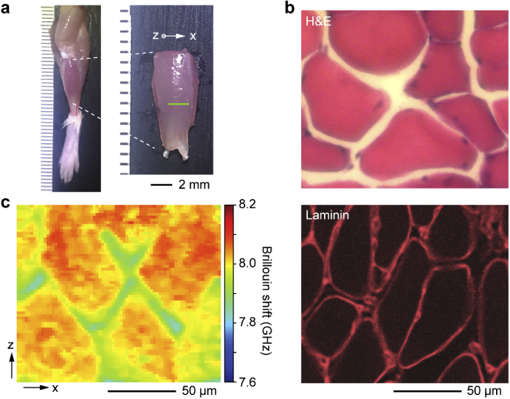
Fig. 1.
Brillouin histology of skeletal muscle tissues. a, Image of the tibialis anterior skeletal muscle obtained from a 6-week old CD-1 female mouse. b, Histology sections of the tibialis anterior skeletal muscle after H&E staining (top) and immunostaining for laminin basement membrane protein (bottom). c, Brillouin shift map along an x-z plane of the intact muscle fascicle.

