Fig. 2.
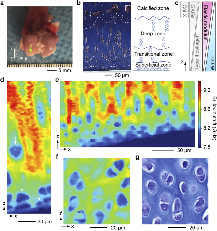
Brillouin histology of articular cartilage tissues from 9-month-old female New Zealand white rabbit. a, Articular cartilage at the femoral head (arrow) was scanned with Brillouin microscopy followed by histological staining for a morphological comparison. b, Masson’s trichrome staining image of a coronal section of the articular cartilage. Dashed lines and an anatomical schematic indicate the four sub-layer zones with chondrocytes. c, Spatial gradients of collagen and GAGs proteins, elastic modulus, and water content [31]. d-e, Brillouin images of an intact articular cartilage tissue in the x-z plane with high spatial resolution (d, 1 µm pixel size; e, 5 µm pixel size). Arrows indicate chondrocytes. ECM in the transitional zone has higher Brillouin shifts than that in the superficial zone. f, Brillouin image in the x-y plane at the transitional zone. Chondrocytes and the stiffer ECM are clearly distinguished. g, Toluidine blue stained image of a x-y cross-sectional tissue section of the transitional zone.

