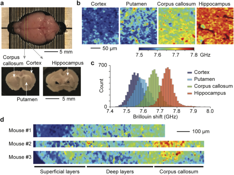
Fig. 4.
Brillouin images of brain tissue slices. a, Coronal brain slices obtained from a 6-week old CD-1 female mouse at -1 and -2 mm from the bregma. b, Brillouin images taken at a depth of 50 µm from the tissue surface at four anatomically distinct sites: cortex, putamen, corpus callosum, and hippocampus. c, Histogram of Brillouin shifts in the four different regions. d, Brillouin maps of brain tissue slices from three different mice taken across the sub-cortical layers and corpus callosum.

