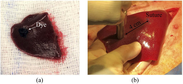
Fig. 1.
Demonstration of the marking strategies implemented for (a) non-survival and (b) survival swine. For non-survival swine, tissue dye was directly applied to the resected liver immediately after each laser application. For survival swine, the laser location was marked by measuring a specific distance (e.g., 3-4 cm) from a suture placed during the first laparotomy.

