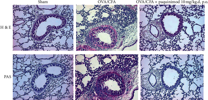
Figure 2.

A representation picture of hematoxylin and eosin (H&E) and periodic acid-Schiff (PAS) staining of lung tissue. Lung sections were obtained from the sham- and OVA/CFA-treated mice with or without paquinimod treatment of 10 mg/kg/day, p.o. Tissue sections were stained with PAS to determine the presence of goblet cells (microscopy image (x200)).
