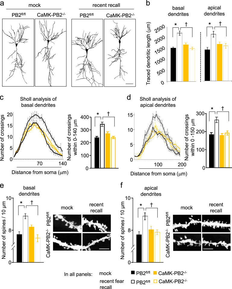
Fig. 4.
Role of Plexin-B2 in fear memory-related remodeling of dendritic complexity and structural plasticity of dendritic spines in hippocampal neurons in vivo. a, b Typical examples of changes in the morphology of Golgi-stained CA1 neurons (a) and quantitative analysis (b) of total length of apical or basal dendrites in CaMK-PB2−/− mice and PB2fl/fl littermates in basal state or at 3 days after fear conditioning (corresponding to recent recall). c, d Cumulative frequency plot of Sholl analysis (left panel) and plot of the average number of dendritic crossings (right panel) in defined distance from the soma of basal (c) and apical (d) dendrites in the treatment groups described above. e, f Typical examples of Golgi-stained dendritic segments (right panel) and quantitative analysis (left panel) of dendritic spine density over basal (e) and apical (f) dendrites of CA1 pyramidal neurons in the treatment groups described above. In all panels, a minimum of 12 neurons per genotype or treatment from at least 3 mice were analyzed. Two-way ANOVA followed by Bonferroni’s test was performed. * represents P < 0.05 when mice with fear conditioning and corresponding mock-treated mice were compared and † represents P < 0.05 when mutant and corresponding wild-type littermates were tested. Error bars represent SEM. Scale bars represent 50 µm (a) and 5 µm (e, f)

