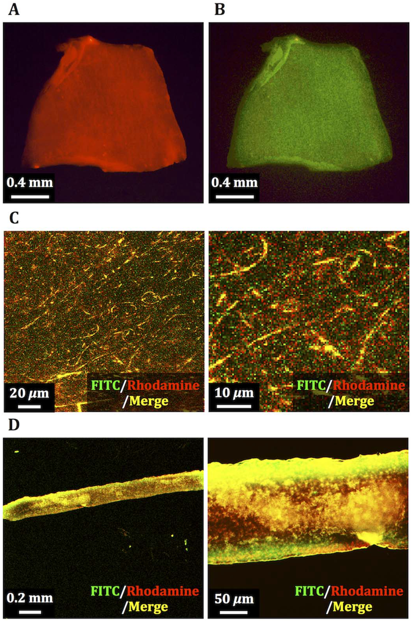
FIGURE 2.

Fluorescence images of PCUU scaffold with FN-G nano-layers composed of rhodamine-labeled fibronectin (Rh-FN) and FITC-labeled gelatin (FITC-G). Fluorescence derived from rhodamine (A) and FITC (B) on a whole scaffold, laser confocal micrographs of fibrous network in a magnified view (C), and cross-sectional images (D) of the PCUU scaffold.
