FIGURE 3.
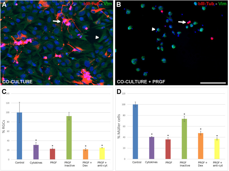
The effect of PRGF on RGCs and Müller cells in co-culture. Control co-cultures of adult pig RGCs and Müller cells (A) and those maintained in the presence of PRGF (B). The RGCs (arrows) were labeled with antibodies against βIII-Tubulin (red) and Müller cells (arrowheads) with antibodies against vimentin (green) and the nuclei were labeled with DAPI (blue). The number of RGCs (C) and Müller cells (D) in the co-cultures relative to the total number of cells in the control condition (100%) are represented in the histograms. RGCs and Müller cells were co-cultured with cytokines (cyt: IL-1β, IL-6 and TNFα), 10% human PRGF (PRGF), heat inactivated PRGF, PRGF with dexamethasone (Dex) and PRGF with antibodies against the cytokines (anti-cyt). Significant differences relative to the control conditions are shown: *p < 0.05. Scale bar = 100 μm.

