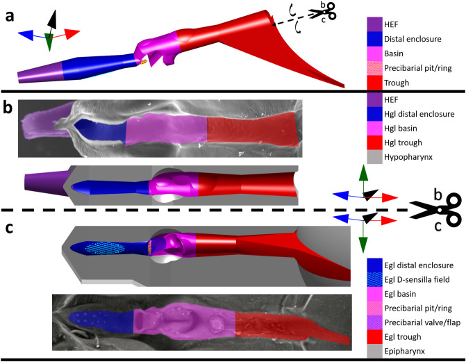
Figure 3.
Close-up segments of the precibarium of the blue–green sharpshooter, labeled with broad terminology. (a) Precibarium 3D model segment. It shows an external view of the walls of the precibarium, as if the rest of the epipharynx and hypopharynx didn’t exist (convex). (b) Hypopharyngeal precibarial walls (colored, concave) in electron microscope image of a segment of the hypopharynx (top, internal) and equivalent view of the 3D model segment (bottom, concave) for comparison. (c) Epipharyngeal precibarial walls 3D model segment (top, mostly concave) and an equivalent electron microscope image (bottom, mostly concave) for comparison. The coordinate axes indicate anterior → posterior, distal → proximal (for the trough), ventral → dorsal, and left → right. The scissors and dotted lines indicate where the 3D model segment in subfigure a would be split to obtain the 3D model segments in subfigures b and c. The associated black curved arrows indicate that the two halves of the split 3D model segment would need to be hinged outward to obtain the views in subfigures b and c. The gray 3D parts of subfigures b and c are semi-abstract representations of the epipharynx and hypopharynx, which serve for comparison with their corresponding electron microscope images. Hgl: hypopharyngeal; egl: epipharyngeal; HEF: hypopharyngeal extension that inserts into the stylet food canal.

