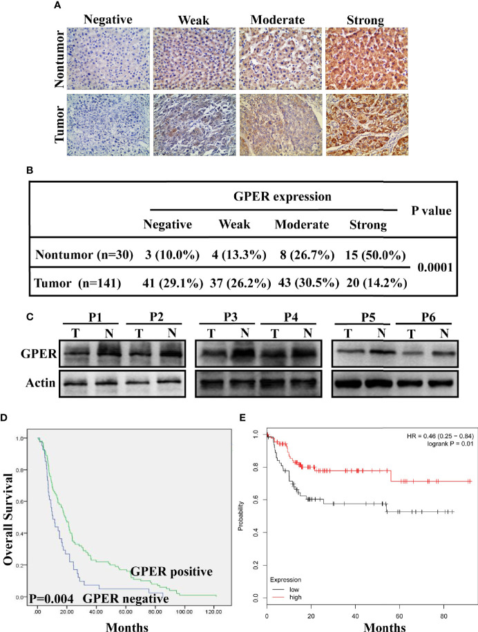
Figure 1.
Clinical significance and prognostic value of GPER expression in hepatocellular carcinoma. (A) GPER expression (negative, weak, moderate, and strong positive staining) in representative non-tumor tissue (top) and HCC tissue (bottom) was analysed using immunohistochemistry (400× magnification, scale bar = 50 μm). (B) IHC analysis revealed significantly different GPER staining between HCC and matched non-tumor tissue (P = 0.0001). (C) Western blot of GPER protein in six pairs of frozen HCC tissue (T1–T6) and matched non-cancerous liver tissue (N1–N6). (D) High GPER level predicts a good overall survival (OS) in patients with HCC (P = 0.004). (E) GPER may be an important protective factor for HCC in Asia (P = 0.01). Data were extracted from a bioinformation database (http://kmplot.com/analysis/).

