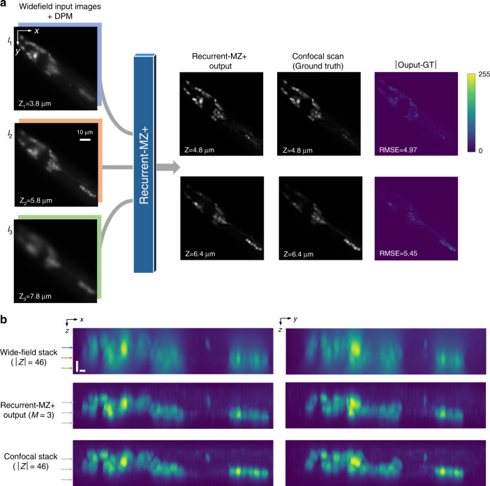
Fig. 8. Wide-field to confocal: cross-modality volumetric imaging using Recurrent-MZ+.
a Recurrent-MZ+ takes in M = 3 wide-field input images along with the corresponding DPMs, and rapidly outputs an image at the designated/desired axial plane, matching the corresponding confocal scan of the same sample plane. b Maximum intensity projection (MIP) side views (x–z and y–z) of the wide-field (46 image scans), Recurrent-MZ+ (M = 3) and the confocal ground truth image stack. Each scale bar is 2 μm. Horizontal arrows in (b) mark the axial planes of I1, I2 and I3. Also see Video S3

