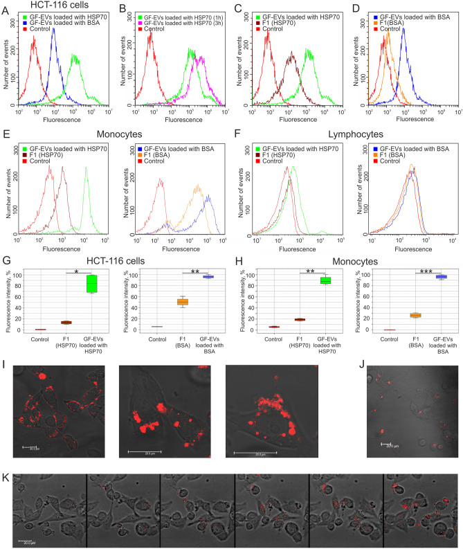
Figure 5.
GF-EV-mediated delivery of exogenous proteins into the human cells analyzed by flow cytometry and confocal microscopy. (A) Incubation of colon cancer HCT-116 cells with GF-EVs loaded with BSA-AF647 or HSP70-AF647 proteins for 1 h. (B) Incubation of HCT-116 cells with HSP70-AF647-loaded GF-EVs for 1 and 3 h. (C,D) Incubation of HCT-116 cells with loaded GF-EVs in comparison with the free HSP70-AF647 or BSA-AF647 proteins for 1 h. (E,F) Incubation of peripheral blood mononuclear cells (E, monocytes and F, lymphocytes) with loaded GF-EVs in comparison with the free HSP70-AF647 or BSA-AF647 proteins for 1 h. (G,H) The relative intensity of fluorescence of HCT-116 cells and monocytes incubated with loaded GF-EVs in comparison with the free HSP70-AF647 or BSA-AF647 proteins. N = 2. Data are presented as means ± SD. *, p < 0.05, **; p < 0.01; ***, p < 0.001 as estimated by one-way ANOVA followed by multiple comparisons Tukey’s post-hoc analysis. (I,J) Micrographs of DLD1 cells co-cultivated with GF-EVs loaded with HSP70-AF647 (I) or BSA-AF647 (J) proteins. Scale bars are 20 µm. (K) Accumulation of fluorescent protein delivered into the recipient DLD1 cells by GF-EVs. Video recording frames were captured every 30 min after adding of loaded GF-EVs to the cells.

