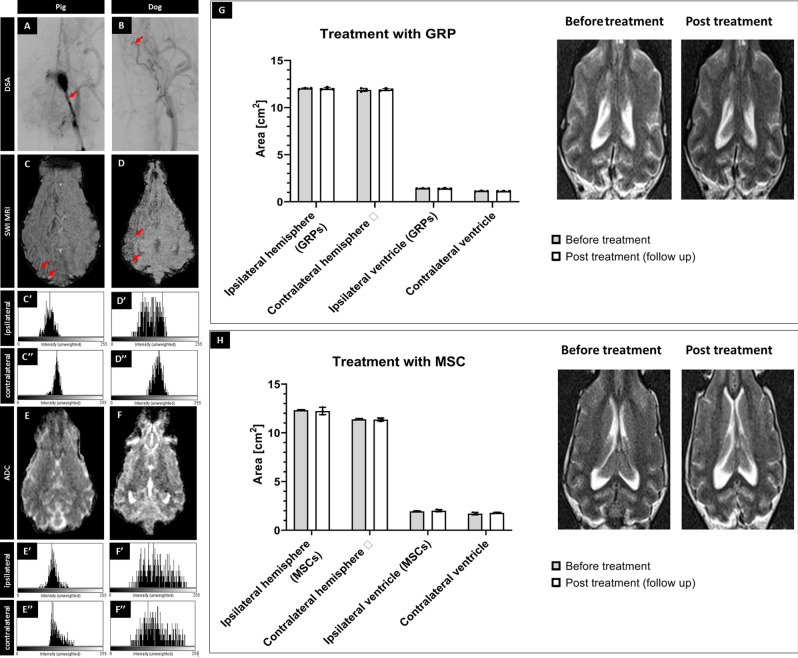
Figure 2.
Safety of the procedures. X-ray evaluation of catheter placement (A,B; arrows indicate placement of the tip of microcatheter). SWI (C,D) with histograms from ipsilateral (C′,D′) and contralateral (C″,D″) hemisphere showing the accumulation of hypointense pixels in the ipsilateral hemisphere. ADC scans (E,F) after pig's and canine's MSC transplantation with histograms from ipsilateral (E′,F′) and contralateral (E″,F″) hemisphere showing any changes in diffusion after cell transplantation. Evaluation of size of hemispheres and ventricles visible on the MRI before versus post-transplantation of GRPs (G) and MSCs (H) showing no differences in size before and after transplantation.

