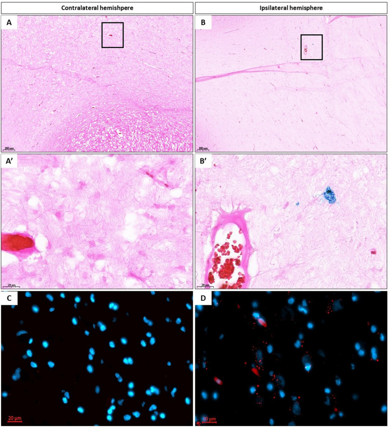
Figure 3.
Histological evaluation. Prussian blue staining of brain of pigs after SPION pMSCs transplantation in contralateral [(A) ×5 magnification, (A′) ×40 magnification] and ipsilateral hemisphere [(B) ×5, (B′) ×40 magnification]. DAPI staining of brain of pigs after SPION pMSC transplantation in collateral [(C) ×40 magnification] and ipsilateral hemisphere [(D) ×40 magnification]. Red spots indicate iron oxide nanoparticles present in pMSCs.

