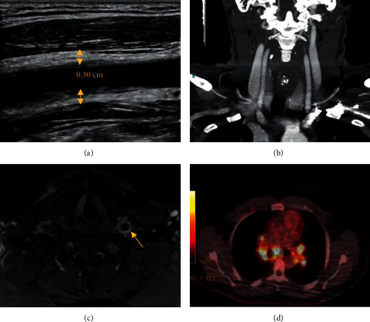
Figure 1.

(a) Longitudinal ultrasound view showing LCC wall thickening (arrows). 0.30 cm relates to vessel wall thickness. (b) Coronal CTA view showing no LCC wall thickening. (c) Axial MRI view showing prominent LCC wall thickening. (d) PET/CT scan showing hypermetabolic mediastinal and hilar lymphadenopathy. ∗Dimensions not to scale. CTA : computed tomography angiography; LCC : left common carotid; PET : positron emission tomography; MRI : magnetic resonance imaging.
