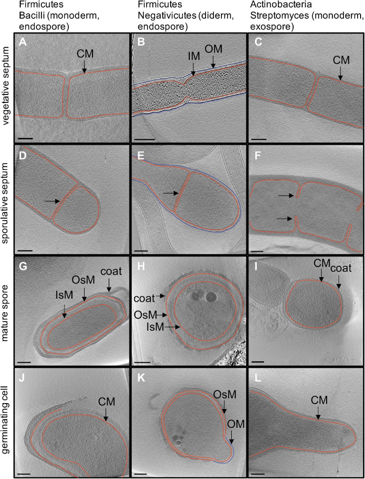
FIGURE 2.
In situ structural detail of sporulation revealed by cryo electron tomography. Each image is a 20-nm slice through a tomogram. Column one (A,D,G,J) outlines major stages of endospore formation in the model organism Bacillus subtilis. Column two (B,E,H,K) outlines endospore formation in Acetonema longum, a diderm Firmicute and member of the Negativicutes. Column three (C,F,I,L) outlines exospore formation in Streptomyces albus. Cytoplasmic (CM) and inner membranes (IM) are shown in red, outer membrane (OM) is shown in blue. The inner and outer spore membranes in Firmicutes (IsM and OsM) are both colored in red to show that they are derived from the CM/IM of the mother cell.

