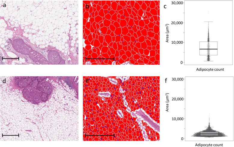
Fig. 2. Digital assessment of breast adipocyte area in two representative patients. Scale bars represent 500 µm.
a A 68-year-old patient with DCIS that developed subsequent iIBC. The breast adipose tissue consists of large adipocytes. b Adipocytes were analysed with the vacuole module v2.1 (HALO software, Indica Labs, Corrales, NM, USA). c Beeswarm plot of the corresponding distribution of adipocyte area with mean adipocyte area and area75th of 7287 and 10493 µm2, respectively. d DCIS surrounded by small adipocytes from a 38-year-old patient. No subsequent iIBC occurred during follow-up. e Corresponding image of analysed adipocytes in HALO software. f Beeswarm plot of the corresponding distribution of adipocyte area with mean adipocyte area and area75th of 2593 and 3274 µm2, respectively.

