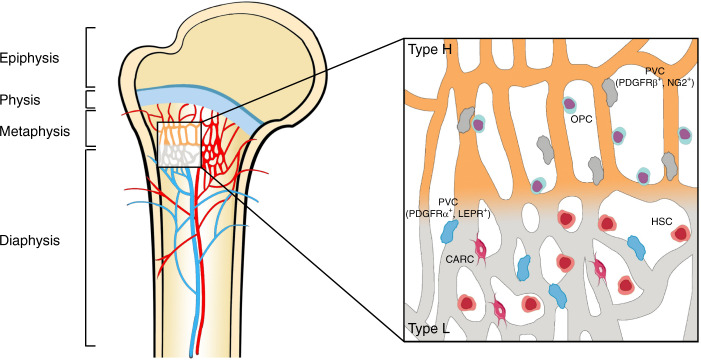
Fig. 2.
Endothelial cell heterogeneity of bone vessels. Distinct capillary subsets with high heterogeneity in the skeletal system can be subdivided into type H and type L endothelium based on morphology, specialization, molecular identity, and functional properties. Type H vessels are primarily distributed around the endosteum region and metaphysis close to the growth plate. They are linearly arranged with distinctive columnar structures and interconnected with new anastomotic loop-like arches at the distal edge. Type L vessels are located with highly branched networks in the bone marrow region of the diaphysis. Type H capillaries are selectively surrounded by Runx2+, collagen 1α+, and Osterix-expressing osteoprogenitors, as well as PDGFRβ- and NG2-expressing perivascular cells. Type L capillaries are predominantly infiltrated by PDGFRα- and LEPR-positive cells as well as CAR cells, which interact with HSCs in the regulation of hematopoiesis. Arteries branch into smaller arterioles and flow into type H vessels in the region of the metaphysis near the growth plate. They then converge into a type L sinusoid network at the interface of the diaphysis and are terminally drained via veins located in the contiguous medullary region

