FIGURE 2.
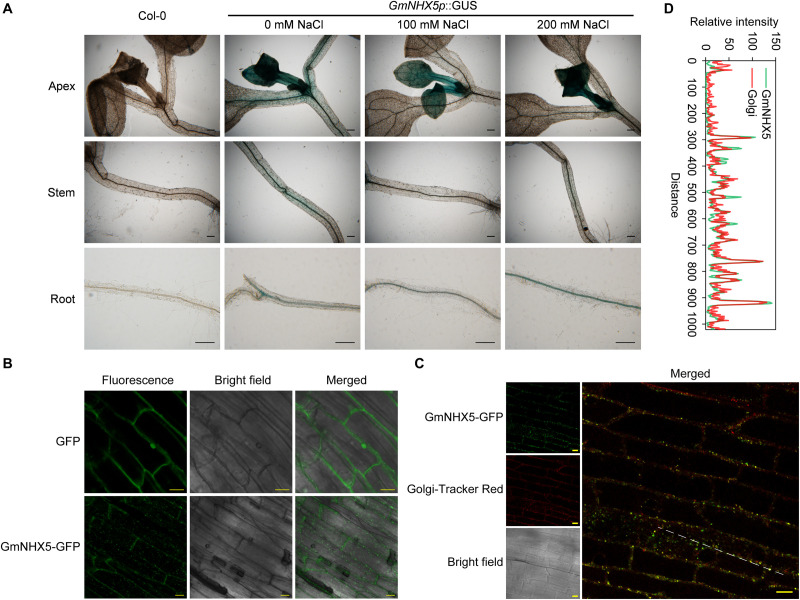
Detection of histological distribution of subcellular localization of GmNHX5. (A) Histochemical analysis of GmNHX5 expression. GUS activity of GmNHX5p:GUS in response to salt stress in transformed Arabidopsis was detected by GUS staining. Five-day-old seedlings were treated with 0, 100, or 200 mM NaCl for 24 h before staining. (B) Fluorescence of GmNHX5-GFP or GFP in Arabidopsis roots. (C) Fluorescence of GmNHX5-GFP and its colocalization with fluorochrome Golgi-Tracker Red. Confocal imaging of the roots of 5-day-old Arabidopsis seedlings transformed with plasmids carrying CaMV 35S:GmNHX5-GFP or CaMV 35S:GFP expression loci. Representative images of three biological replicates are shown. Bars = 200 μm in panel (A), Bars = 10 μm in panels (B,C). (D) Line profile was used to illustrate colocalization between GmNHX5-GFP and Golgi-Tracker Red along the dotted line in panel (C). Green and red lines indicate GmNHX5-GFP and Golgi-Tracker Red fluorescence profiles, respectively.

